Photoelectron emission microscopy (PEEM)
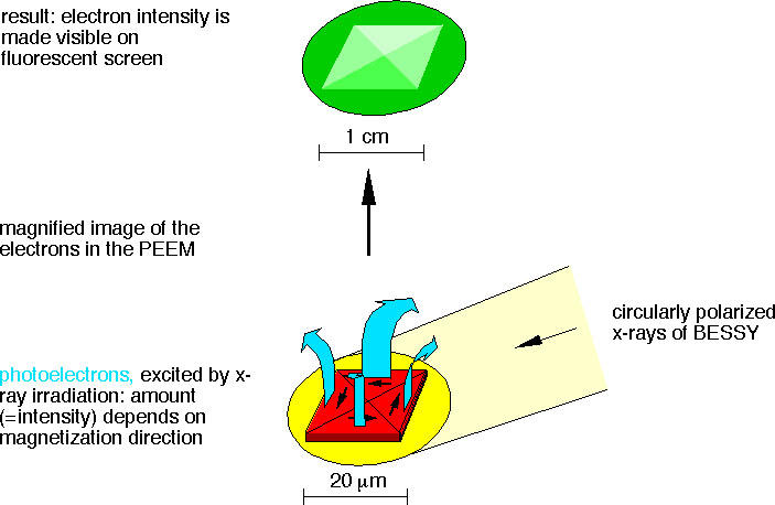
 PEEM is an electron-optical parallel imaging technique. Electrons emitted from the surface of a specimen after absorption of photons are used to obtain a laterally resolved image of the surface. The intensity in the image represents the local electron photoyield, and thus the local photon absorption cross section.
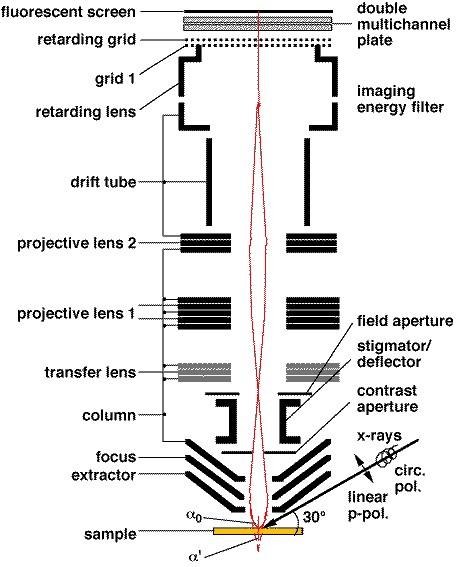
The image shows a sketch of an electrostatic instrument. Electron lenses create a magnified image of the photoexcited electrons emitted from the sample, similar to an electron microscope (basic resolution: 20 nm). The image is projected onto a fluorescent screen.
Magnetic properties on the sub-micrometer scale can be imaged taking advantage of the XMCD effect. For excitation at elemental absorption edges with circularly polarized x-rays, the electron intensity depends on the relative orientation of (local) magnetization direction and light helicity, thus generating magnetic contrast.
(Click on images to get an enlarged view.)
 |
 |
Photoelectron emission microscopy (PEEM) and x-ray magnetic circular dichroism (XMCD) combine lateral resolution with element-sensitive magnetic contrast, which is essential for investigating multilayer systems.
Time-resolved PEEM
To investigate magnetization dynamics in “real time”, the sample is excited by short magnetic field pulses. The response of the sample to the field pulses is stroboscopically investigated by magnetic microscopy.
The figure sketches the principle of stroboscopic imaging of magnetization dynamics by time- and layer-resolved XMCD-PEEM. Here, microstructures are deposited onto a stripline, which is connected to a photoconductive switch. By focusing a fs laser onto the switch, a current pulse through the stripline is generated and thereby a magnetic pulse in the vicinity of the structures. Synchronization of the magnetic pulses to the x-ray pulses of the BESSY single bunch operation mode (probe) results in a stroboscopic pump–probe experiment.
Alternatively to field pulses triggered by a photoconductive switch, samples can be also excited by ultrashort laser pulses.
