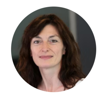
Physikalisches Kolloquium: Vortrag im Rahmen eines Symposiums zur Biophysik - Prof. Paola Borri "Shedding new light on cells with coherent optical microscopy"
Cardiff University School of Biosciences
Optical microscopy is an indispensable tool that is driving progress in biology and is still the only practical means of obtaining spatial and temporal resolution within living cells and tissues. In this context, staining samples with fluorescent labels provides a highly sensitive and specific method of visualizing biomolecules. However, fluorescence microscopy has limitations including sample manipulation and staining artifacts, fluorophore photobleaching and associated phototoxicity. Therefore, much effort has been devoted to developing alternative optical microscopy techniques which are non-perturbing, photostable, and in turn offer quantitative capabilities unavailable with fluorescent methods.
Coherent Raman Scattering (CRS) microscopy has attracted increasing attention as a powerful multiphoton microscopy technique which overcomes the need for fluorescent labelling and yet retains biomolecular specificity and intrinsic 3D resolution. Over the past 15 years, our laboratory has developed and demonstrated a range of CRS microscope set-ups featuring innovative excitation/detection schemes. Our second-generation coherent anti-Stokes Raman scattering (CARS) instrument is based on a pulsed 5fs Ti:Sa laser source capable of exciting a wide vibrational range (from 1000cm-1 to 3500cm-1) which allows us to perform hyperspectral microscopy, whereby a vibrational spectrum is obtained for each spatial voxel. This in turn has led to the development of quantitative chemical imaging algorithms to represent the hyperspectral dataset as a superposition of Raman spectra and concentration maps of individual chemical components (e.g. proteins, lipids, DNA) in physically eaningful units.
With this technique, we have measured the 3D spatial distribution of lipid droplets in live mouse oocytes and early embryos and elucidated the link between this distribution and the use of fatty-acids in the egg metabolism. We have also quantitatively measured volumetric concentrations (and in turn dry masses) of lipids, proteins and DNA in 3D during cell division. Furthermore, we have shown that CARS can be used to detect single non-fluorescing nanodiamonds in cells for the first time. Recently, we have applied the technique to visualise lipid partitioning in single planar membranes bilayers exhibiting liquid ordered (Lo) and disordered (Ld) domains. We also developed an interferometric CRS set-up, which offers background-free chemically-specific image contrast, shot-noise limited detection, and phase sensitivity, enabling topographic imaging of interfaces.
Complementary to CRS, we have invented and developed a four-wave mixing (FWM) interferometry technique, whereby single small (≥5nm radius) gold nanoparticles are detected background-free, owing to their specific and photostable nonlinear plasmonic response, even inside highly scattering/auto-fluorescing biological specimens including multicellular organs. The set-up enables correlative FWM/confocal fluorescence imaging, which we used to study the fate of nanoparticle-biomolecule-fluorophore conjugates and their integrity inside cells. The technique lends itself to tracking single particles with nanometric position localisation precision in 3D at high speed (sub-ms), opening the exciting prospect of unravelling single-particle trafficking within complex 3D architectures over virtually unlimited observation times. Notably, gold nanoparticles are electron dense and widely used probes in electron microscopy. By exploiting FWM, we recently demonstrated a novel correlative light-electron microscopy approach using gold nanoparticles as the same probe visible inside cells in both imaging modalities, bringing high correlation accuracy without the need for additional fiducial markers.
Besides nonlinear optical techniques, we are also working on quantitative differential interference contrast microscopy to measure the thickness of individual lipid bilayers with sub-nm precision, and have recently developed a widefield interferometric reflectometry method, aimed at monitoring single protein-lipid membrane interactions with unprecedented sensitivity.
I will present our latest progress with these techniques and their applications to bio-imaging.
Zeit & Ort
30.06.2023 | 15:00 c.t. - 17:00
Hörsaal A (1.3.14)
Weitere Informationen
Gastgeberin: Prof. Dr. Cecilia Clementi
